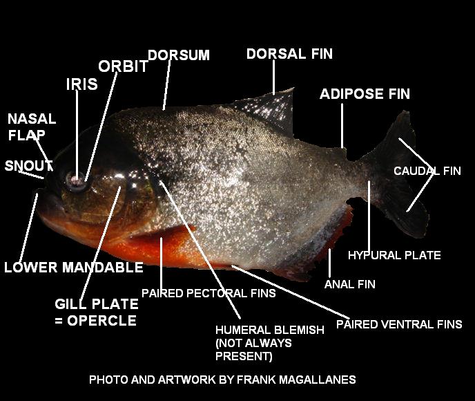
Body Parts of a Piranha, what is the fish made of?
All photos and illustrations by Frank Magallanes unless otherwise noted.
UNDER REVISION - WITH NEW PHOTOS AND INFORMATION ADDED SOON - Work started March 23, 2012
temporary work completed 21 Apr 2012
INTRODUCTION
The information presented here will be generalized. However, I will be giving some specific information in the way of drawings and some images, for in depth studies by students. Much of the work on the bone structure of characins was taken from Dr. Stanley Weitzman in his descriptive work entitled: Osteology of Brycon meeki (1962). What I have done here is taken portions of the citation and incorporated it to fit piranha morphology. Readers should not assume that this page is endorsed by Dr. Weitzman or any other Systematicians. However, I have taken pains to ensure the work presented here is as accurate as humanly possible. The web page was critiqued by a friend and PhD in ichthyology who is familiar with piranha bone structure. His supervision is without a doubt, helpful in creating this page on piranhas.
|
Dr. S. Weitzman, a Hobbyist, Scientist, Systematician I truly admire. |

Miriam L. Zelditch and William L. Fink
Abstract
Piranhas, like many teleosts, change their diets on both ontogenetic and Phylogenetic time scales. Prior studies have suggested that pervasive morphological changes in body form on a Phylogenetic time scale may be related to changes in diet, but previous reports have found little shape change in piranhas on an ontogenetic time scale. We re-examine the post-transformational allometry of body form in one piranha, Pygocentrus nattereri (Kner), using the method of thin-plate splines decomposed by their partial warps. We find substantial evidence of allometry, primarily elongation of the mid-body relative to the more anterior and posterior regions, elongation of the postorbital and nape regions relative to the more anterior head and posterior body, and deepening of the head relative to the body. In addition to these pervasive changes throughout the body, there are some that are more localized, especially elongation of the postorbital region relative to eye diameter and snout, and an even more localized elongation of the snout relative to eye diameter. Initial dietary transitions are associated with changes in head and jaw proportions, but rates of shape change decelerate through growth, so that the final transition to a diet increasingly dominated by small whole fish appears associated with change largely in overall body size.
The Head
Piranhas (genus Pygocentrus) are variable in head shape (slope) dependant on age and food. For that reason alone some confusion exists among hobbyist that collect piranhas. Historic literature pertaining to classification used vague terms like; "lower jaw very heavy." Piranhas brought into the hobby as wild caught present another issue. The genetics of in-breeding is not as prevalent as those of aquarium raised species where constant mixing of same related stock replenishes the genetic pool causing problems in identification and skeletal structure. I caution hobbyist to be wary of fish offered for sale that are declared different than ordinary similar species. This feature is in fact very common dependant on the geographical location and the consistency of the piranhas diet.
|
|
|
 |
|
As the fish develops the slope area will become more convex until the blunt-headed shape becomes prominent. Not all piranhas of genus Pygocentrus develop this convex shape. But it appears its diet is the main governing force on what the head shape will be like. Fink et al., found no significant differences between southern and Amazonian populations in these features. Snout length does appear to be short in large individuals from the southern populations but is not significantly different from Amazonian populations.
|
Examples of wild caught P. nattereri. Note the variance of body and head shape. The Piranha Book - G.S. Myers |

The widespread P. nattereri has apparently two forms, a northern form in the Amazon basin, and a southern form previously described as P. ternetzi described by Steindachner (1908). Fink and Zelditch (1997) could not find reliable characteristics to distinguish between the two, and consider P. ternetzi as a nonlinear cline (varying body shape) of P. nattereri. These are often sold as "Yellow Emperor Piranhas" because of the head shape and coloration, but are nothing more than southern populations of P. nattereri.
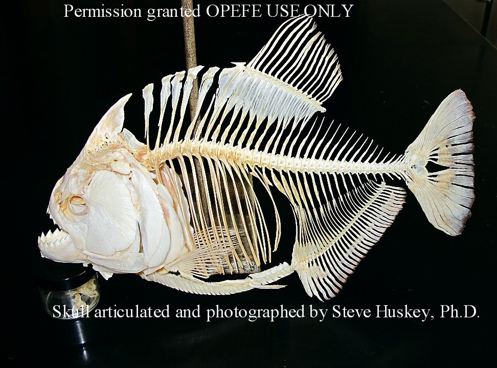
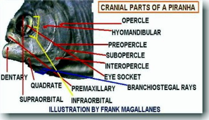
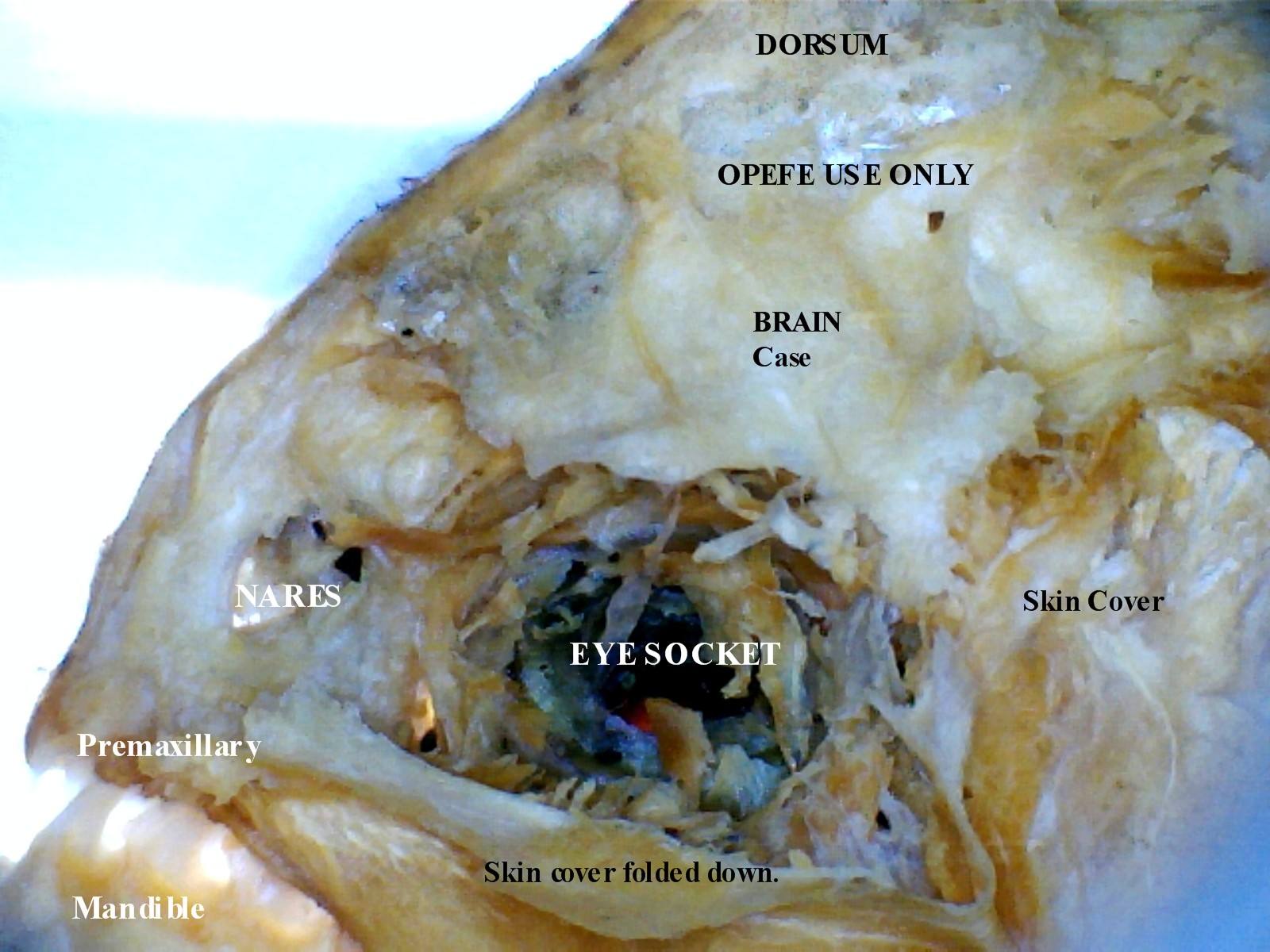
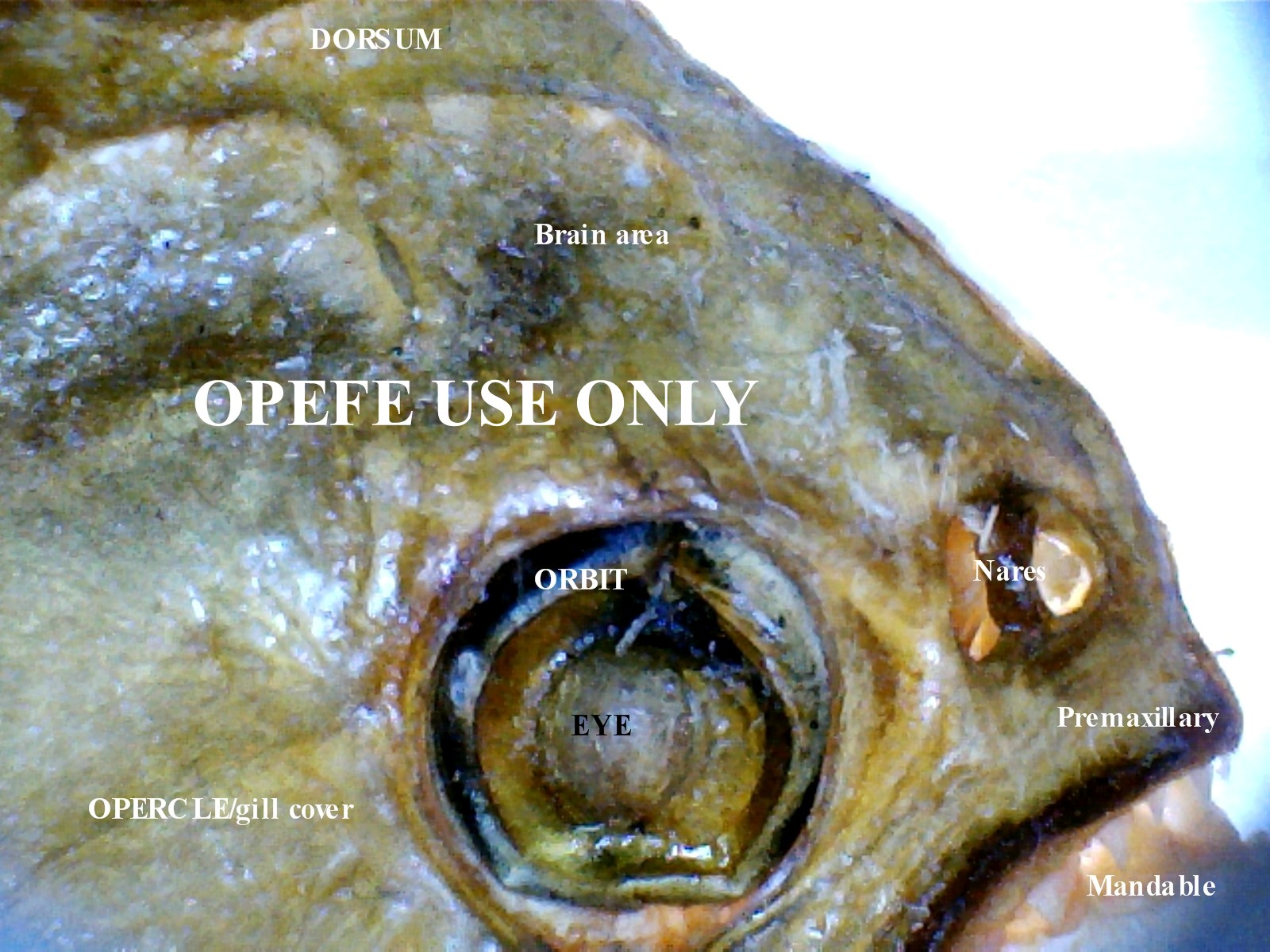
The skull (or syncranium) of fishes are divided into two basic units, neurocranium (brain case) and branchiocranium. The upper jaw, maxillary and premaxillary bones are attached to the neurocranium. The opercular series is conected to the suspensorium and the hyoid complex to form the gill chamber. This chamber works in unison to allow water to pass through the mouth and ventilate the gills. The gill arch is composed of five bones. Starting at the ventral anterior end of the arch the parts are brasibranchial, hypobrancial, ceratobrancial, and interbrancial. Nearly all fishes have these gills which are composed of cartilaginous or bony tubercles, called gill rakers, which are attached to the inside of the gill arches. The function of the gill rakers has two purposes; protect the delicate gill filaments from foreign particles or to strain out planktonic organisms to be used as food. The blood to these gills are aerated by the rhythmical intake of water of the oral cavity (mouth) and its expulsion through the gill clefts. This then passes the gill filaments. The gill filaments absorb the oxygen and go out as waste byproducts of dissolved gases. The membrane (which can be seen when the fish mouth is open) acts as a valve to prevent water from moving in the wrong direction. If water conditions are bad this can effect the fishes ability to take in oxygen and cause too much carbon dioxide in the blood resulting in labored breathing.
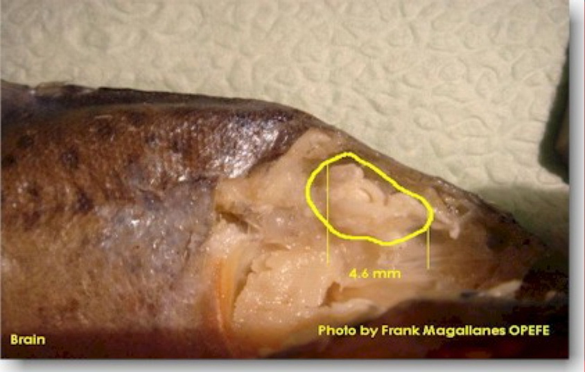
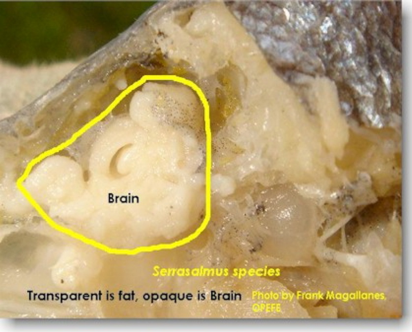
The Brain
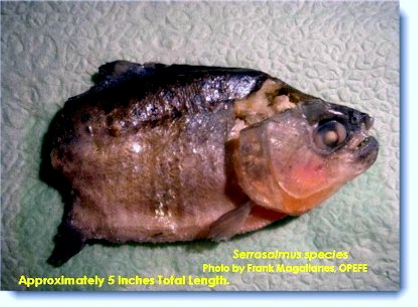
Example below is of a split Serrasalmus sanchezi showing the partial brain case and brains.
|
Serrasalmus |
Serrasalmus |
Closeup of brain |
Pygocentrus |
Pygocentrus |
The body
 Evolution of shape - In
a recent analysis of piranha shape evolution, juveniles were found to have a
greater diversity of body shapes than adults. Although it is commonly assumed
that early ontogenetic stages are more conservative than those that follow
later, diversity actually drops when these fishes move from juvenile stages to
adults. This increase in juvenile diversity is the result of a highly modified
juvenile shape in one of the lineages included in the study. (Fink 1996) Another
factor being studied by today's Systematicians is how these developmental
pathways evolved. Thus giving rise to the diversity and evolution of body shapes
seen in piranhas today.
Evolution of shape - In
a recent analysis of piranha shape evolution, juveniles were found to have a
greater diversity of body shapes than adults. Although it is commonly assumed
that early ontogenetic stages are more conservative than those that follow
later, diversity actually drops when these fishes move from juvenile stages to
adults. This increase in juvenile diversity is the result of a highly modified
juvenile shape in one of the lineages included in the study. (Fink 1996) Another
factor being studied by today's Systematicians is how these developmental
pathways evolved. Thus giving rise to the diversity and evolution of body shapes
seen in piranhas today.
 The body shape is very important in
identifying the type of piranha you own, particularly if you wish to
differentiate from Pygocentrus and say, Serrasalmus species.
Generally, Pygocentrus species when adult, are stocky, broad forehead and
snout and the under jaw gives the entire head and jaw the appearance of a
bulldog. All species in the genera are high-backed. A look at the Serrasalmus
species on the other hand, they generally have a more pointed snout and jaw,
and a very compressed body. They all possess the belly saw or serrae. Generally,
male piranhas are more slender, but even in large groups, it is not always
possible to detect which are which. In the home aquarium, you may have some luck
in determining the sexes if they breed for you. Then you will be able to tell
positively which are male and female if you pay close attention. Otherwise, it's
extremely difficult to tell.
The body shape is very important in
identifying the type of piranha you own, particularly if you wish to
differentiate from Pygocentrus and say, Serrasalmus species.
Generally, Pygocentrus species when adult, are stocky, broad forehead and
snout and the under jaw gives the entire head and jaw the appearance of a
bulldog. All species in the genera are high-backed. A look at the Serrasalmus
species on the other hand, they generally have a more pointed snout and jaw,
and a very compressed body. They all possess the belly saw or serrae. Generally,
male piranhas are more slender, but even in large groups, it is not always
possible to detect which are which. In the home aquarium, you may have some luck
in determining the sexes if they breed for you. Then you will be able to tell
positively which are male and female if you pay close attention. Otherwise, it's
extremely difficult to tell.
|
|
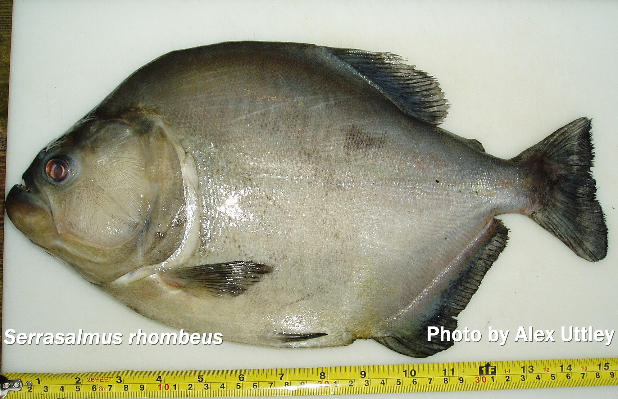
The body is built for its environment. Those that are torpedo-shaped or semi-elongated are normally found in fast flowing waters. Those that are somewhat stocky, generally are found in slow-moving waters or stagnant waters. The more compressed the species is, the more likely to find it inhabiting submerged roots of trees and plants. Some species like Serrasalmus rhombeus, are found in very deep fast flowing waters near rapids, their powerful deep bodies are built for that environment. Others like Pygopristis denticulata are found in ponds or lakes where the current is not so strong.
Sense Organs and how it applies to Piranhas
Sense organs acquaint fishes with their immediate environment. The simplest of these are the cutaneous sense organs. In piranhas, this is represented by the lateral line which is an embedded tube, extending along the sides. The functioning of the lateral line system enable fishes to detect low-frequency vibrations in the water.
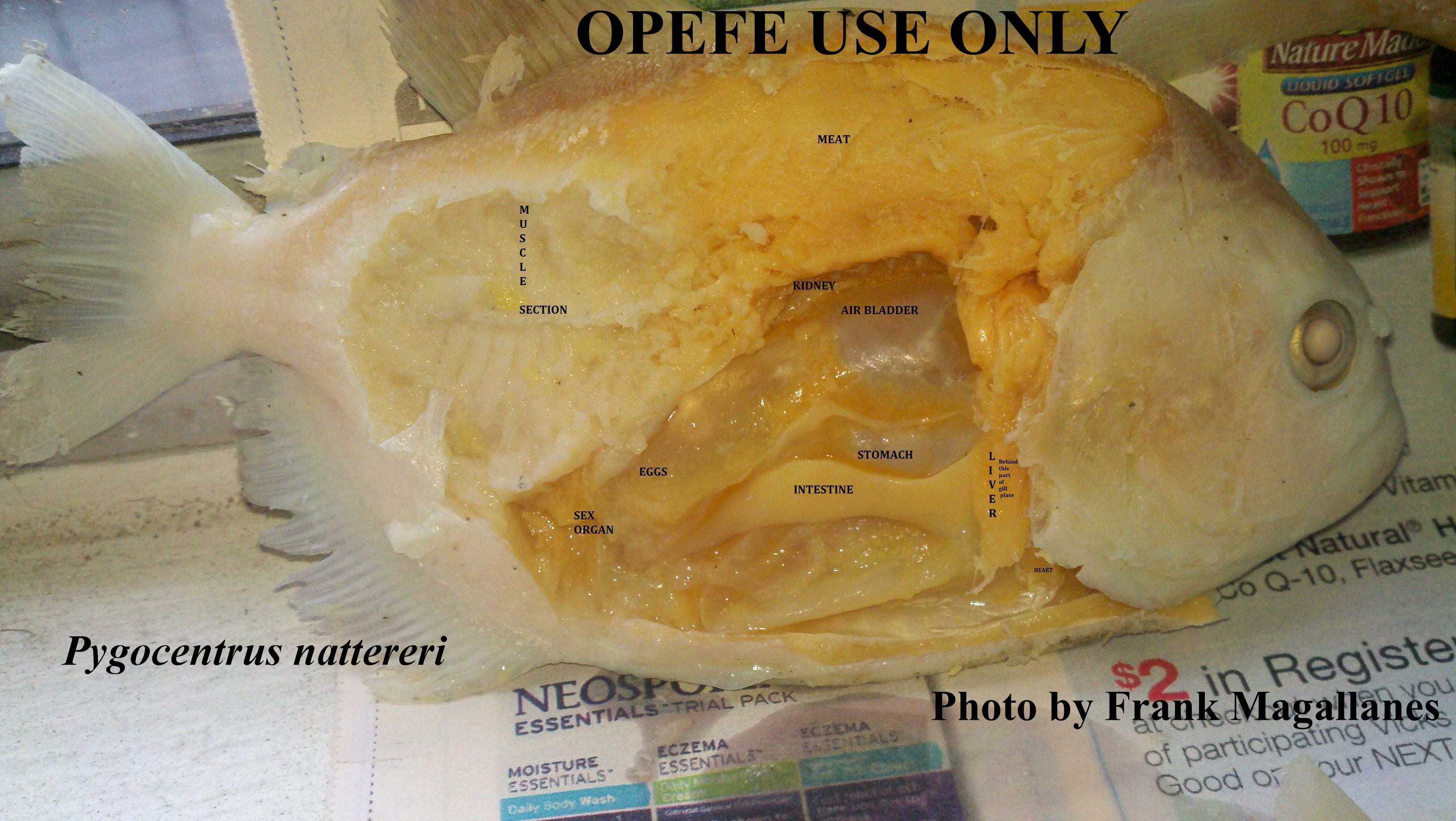
Internal and Sex organs
|
|
|
|
|
|
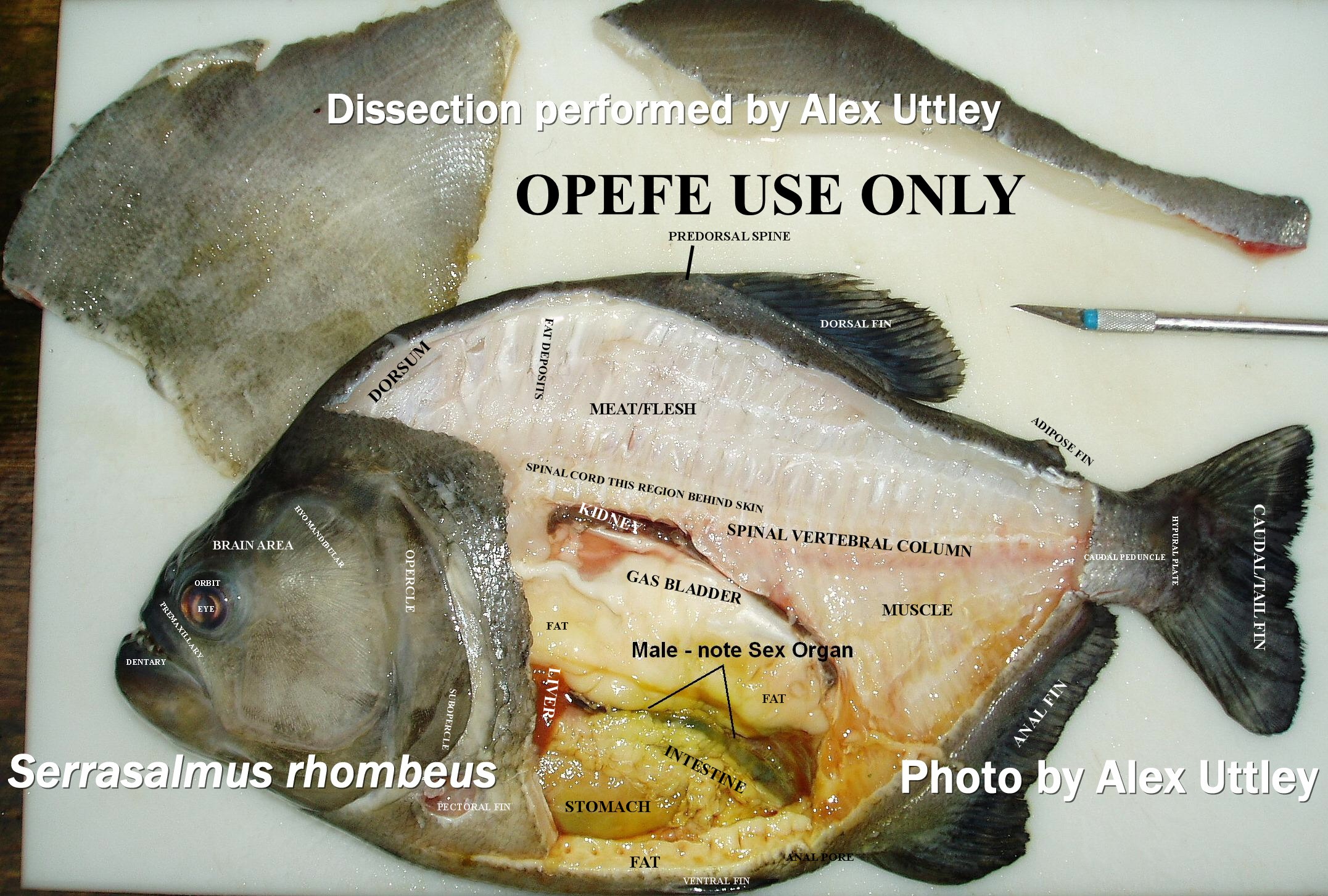
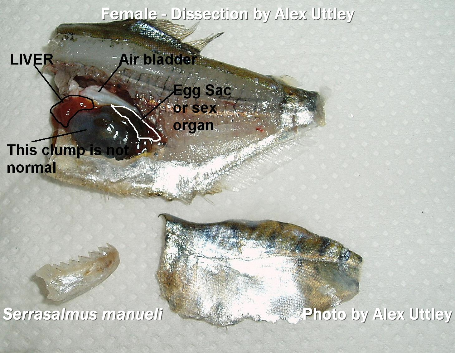
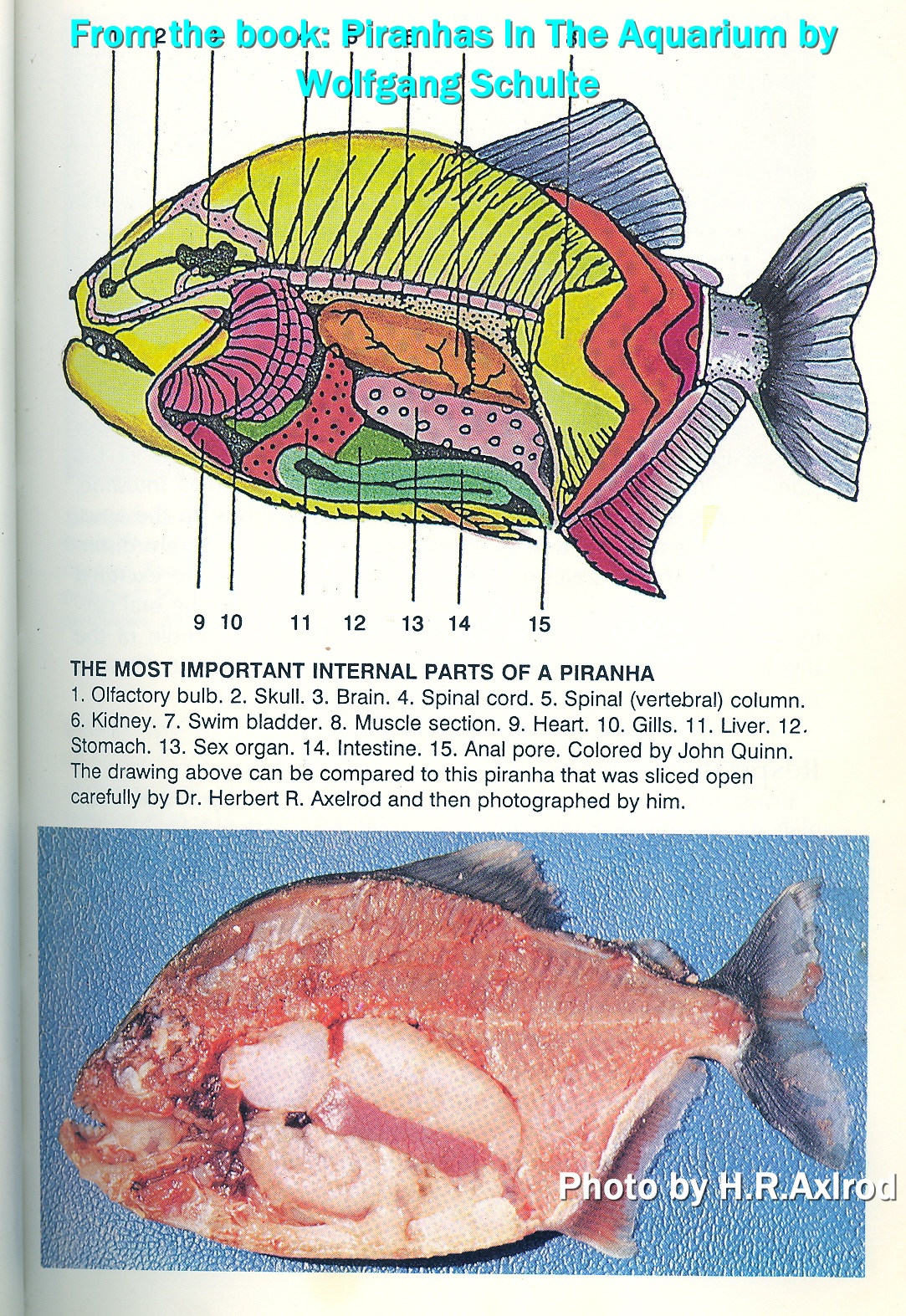
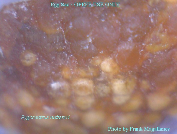
While only two (2) piranha species show sexual dimorphism, the internal organs show something much more different. Unfertilized eggs can be seen in the examples above. The John R. Quinn & Herbert R. Axelrod artwork and photography show the areas to be examined and what the organs are. Note the H. Azuma photography 2nd to last on right.
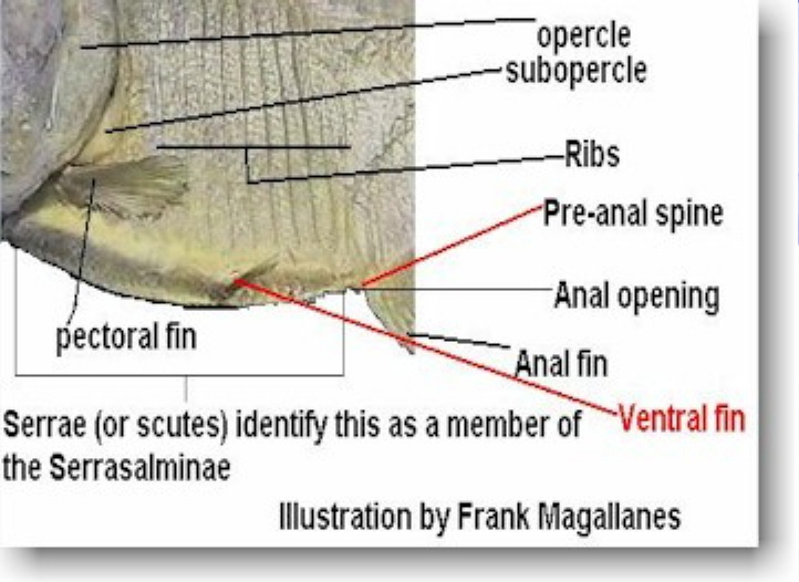
Serrae and scutes
|
|
|

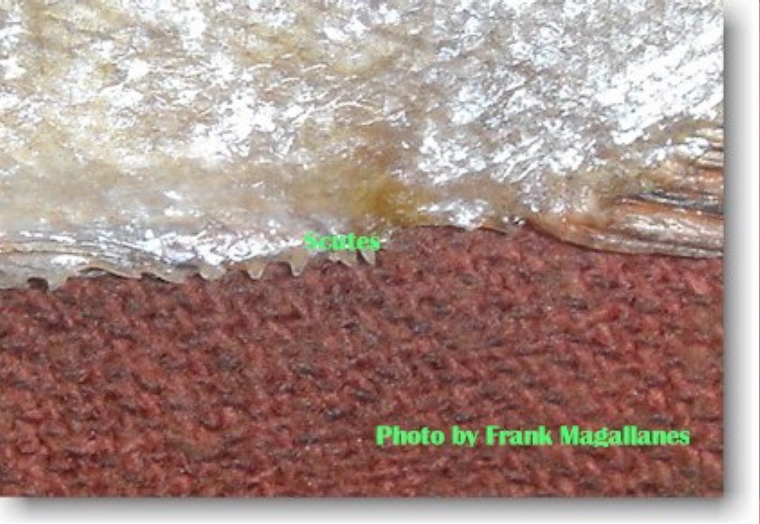
Scutes
and serrae are small bones that resemble a saw-blade and help identify certain
piranha or pirambeba. Some like S. sanchezi are prominent (see photo at
left). A scutes is a bony type of projection. Often is a modified scale.
Presence or absence, location, count, and type of scutes is very useful in
identifying certain species of fish.
|
|
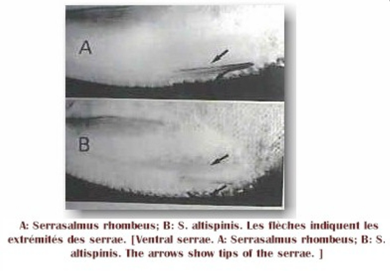
Some species like S. altipinis and S. rhombeus (above photo) have unique features. S. altispinis scutes (lower top photo) are more prominent and form the description for the species which helps differentiate it from its closest relative, S. rhombeus (top photo).
 Some like S. sanchezi,
the serrae are large and pronounced in comparison to those two species and those
found in the spilopleura complex group, but often seen with the Pristobrycon-Humeralis
group. Serrae are important to scientists because it helps define the family
these fishes belong to; Serrasalminae, which is serrated salmon family.
Some like S. sanchezi,
the serrae are large and pronounced in comparison to those two species and those
found in the spilopleura complex group, but often seen with the Pristobrycon-Humeralis
group. Serrae are important to scientists because it helps define the family
these fishes belong to; Serrasalminae, which is serrated salmon family.
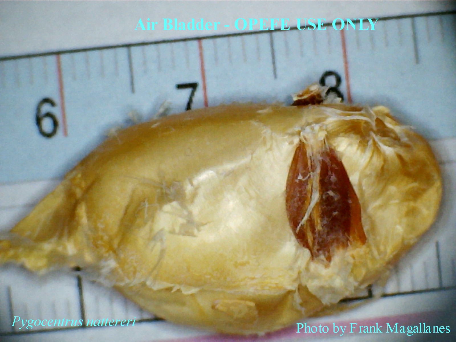
Air Bladder
Generally, piranhas of the different species have unique air bladders. Some are double chambered others long or short.
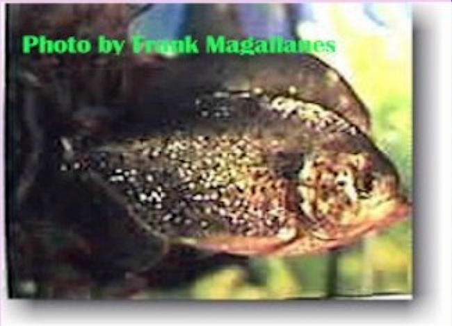
Humeral blotch
 The humeral blotch also commonly called humeral spot
is a non-fixed character found prominently in some species of Piranhas (i.e., S.
manueli, P. cariba, Pristobrycon sp.). The humeral blotch may
be indicative of a species such as Pygocentrus cariba and may be part of
the fishes early development, but later fading to a smaller triangular spot or
none at all. This however is not fixed even within that population though it is
one of the characters listed for the species. It is basically a recognition
symbol between species. Some fishes like S. eigenmanni have the spot
fixed in some localities, in others it is absent in majority of specimens. This
character alone does not erect a species as solely distinctive, since it is
variable among so many populations of same species. Some present work can be found among cichlid data collected by
researchers where the humeral spot is present in same species, but in others
not. There is no concrete explanation for either the piranhas or the cichlids
why the humeral blotch is variable among same species. .
The humeral blotch also commonly called humeral spot
is a non-fixed character found prominently in some species of Piranhas (i.e., S.
manueli, P. cariba, Pristobrycon sp.). The humeral blotch may
be indicative of a species such as Pygocentrus cariba and may be part of
the fishes early development, but later fading to a smaller triangular spot or
none at all. This however is not fixed even within that population though it is
one of the characters listed for the species. It is basically a recognition
symbol between species. Some fishes like S. eigenmanni have the spot
fixed in some localities, in others it is absent in majority of specimens. This
character alone does not erect a species as solely distinctive, since it is
variable among so many populations of same species. Some present work can be found among cichlid data collected by
researchers where the humeral spot is present in same species, but in others
not. There is no concrete explanation for either the piranhas or the cichlids
why the humeral blotch is variable among same species. .
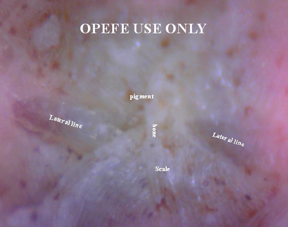
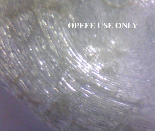
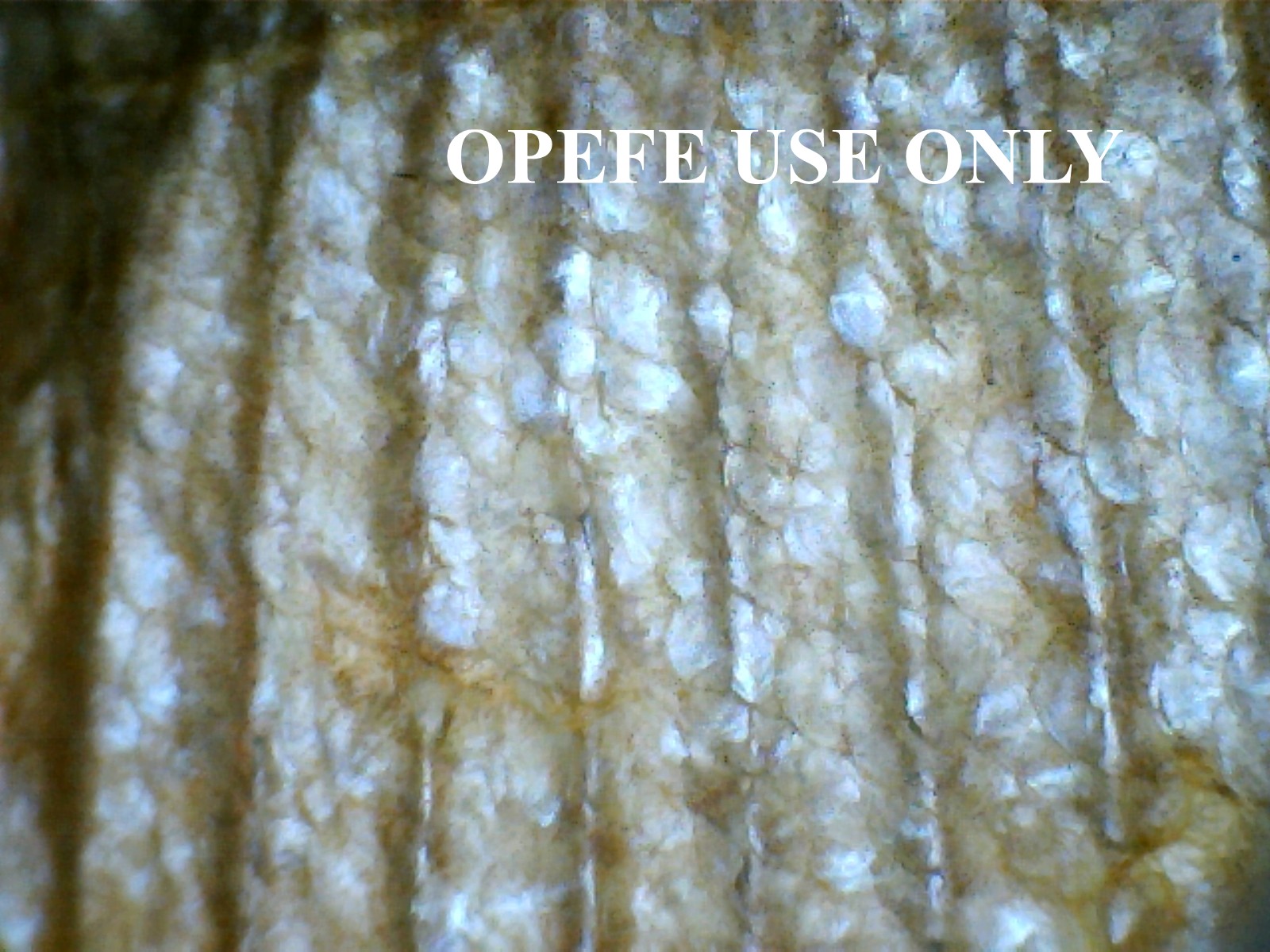
Scales and their coloration
Systematicians use scale counting (vertical and horizontal scale rows) to reach a formula and desired scale count. Piranha scales are circuli similar to tree growth rings. Most piranha hobbyists are unaware that it is not the scale that produces the color themselves, but the pigment-containing cells or what is known as chromatophores. These cells are in the fish skin where the scales are embedded. Sunlight stimulates these cells to make them glimmer in an array of different colors or if they are clustered together to make one color. An example of this is the red throat of Serrasalmus sanchezi. This also helps species recognize each other. The cells may also impact the eyes giving the same species some distinctiveness like those in the ruby red eyes of S. rhombeus.
|
complete scale view |
Magnified and viewed |
Under microscope |
How scales are aligned. |
 Color is a plastic character that can vary wildly within
the geographic range of these fish. There are several cases of regional
differences, because of environmental conditions or other factors.
Piranhas in the aquarium lack this ability to sustain their wild caught colors
for any length of time. There are exceptions, but very few examples. The belly
colors ranging from red-orange-yellow is common to the entire genus of Pygocentrus
depending on geographical location. While "color of life" is a
good marker in some situations to identify a species it is not a good character
to determine a particular species based on that alone. At rest, the fishes color
become bland or gray during sleep hours. Light, as discussed above stimulate the
color of the fish as well as the type of substrate being used. Bright lights and
light color can wash out a fishes color, too dark of a color and the fish will
be dark gray to black. Sunlight brings out the best color in fishes, however in
the aquarium it can cause an algal bloom. So proper lighting must be obtained
via florescent tubes that are rated for color and intensity. Other conditions
that can effect color in fishes is the amount of stress the fish is being
subjected to. Illness can make a fish appear to be in breeding condition (dark).
Poor water conditions can also make the fish become dull and colorless. Age is
another factor that is often overlooked. Old fish generally become darker and
less intense in color.
Color is a plastic character that can vary wildly within
the geographic range of these fish. There are several cases of regional
differences, because of environmental conditions or other factors.
Piranhas in the aquarium lack this ability to sustain their wild caught colors
for any length of time. There are exceptions, but very few examples. The belly
colors ranging from red-orange-yellow is common to the entire genus of Pygocentrus
depending on geographical location. While "color of life" is a
good marker in some situations to identify a species it is not a good character
to determine a particular species based on that alone. At rest, the fishes color
become bland or gray during sleep hours. Light, as discussed above stimulate the
color of the fish as well as the type of substrate being used. Bright lights and
light color can wash out a fishes color, too dark of a color and the fish will
be dark gray to black. Sunlight brings out the best color in fishes, however in
the aquarium it can cause an algal bloom. So proper lighting must be obtained
via florescent tubes that are rated for color and intensity. Other conditions
that can effect color in fishes is the amount of stress the fish is being
subjected to. Illness can make a fish appear to be in breeding condition (dark).
Poor water conditions can also make the fish become dull and colorless. Age is
another factor that is often overlooked. Old fish generally become darker and
less intense in color.
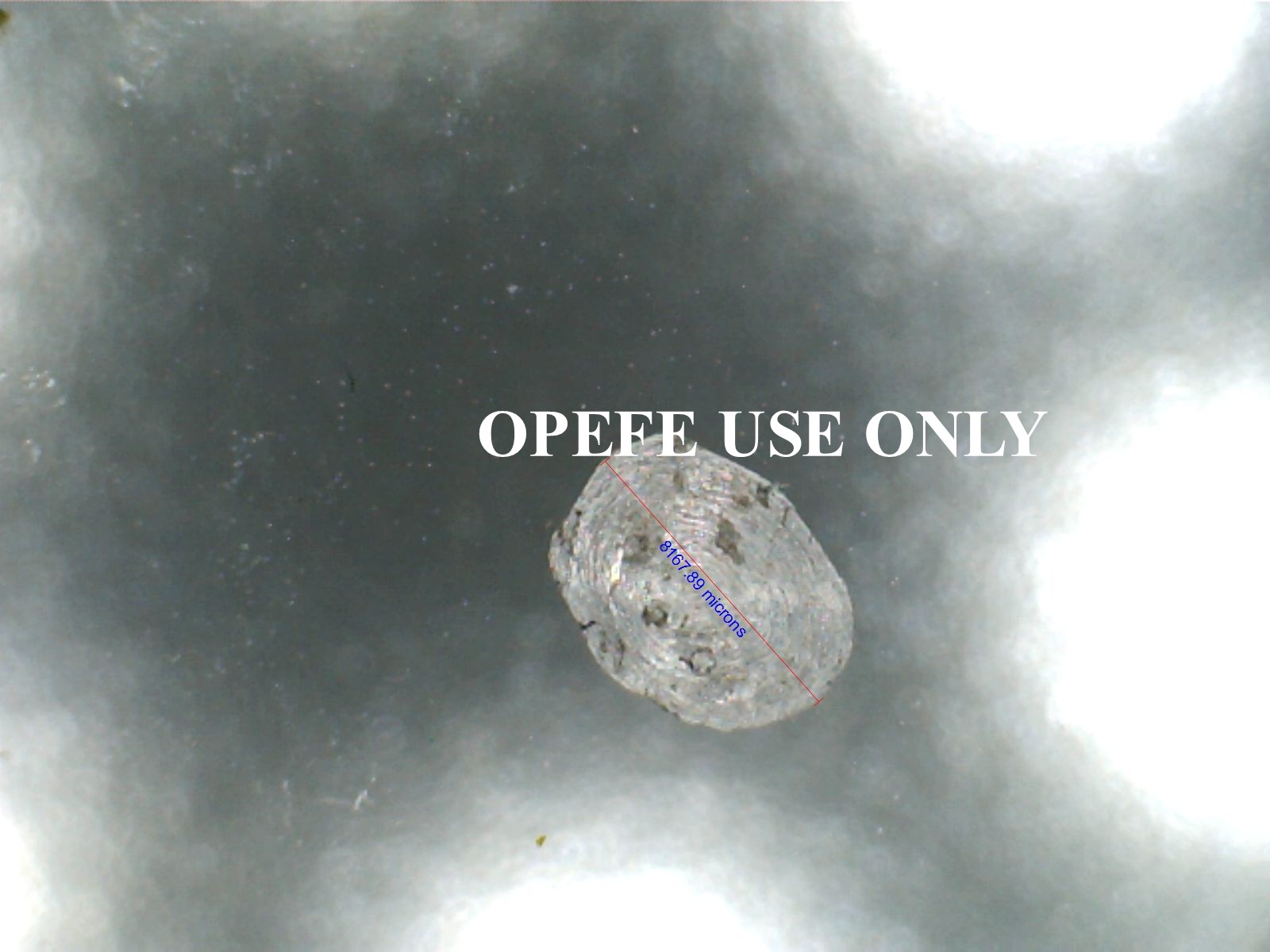
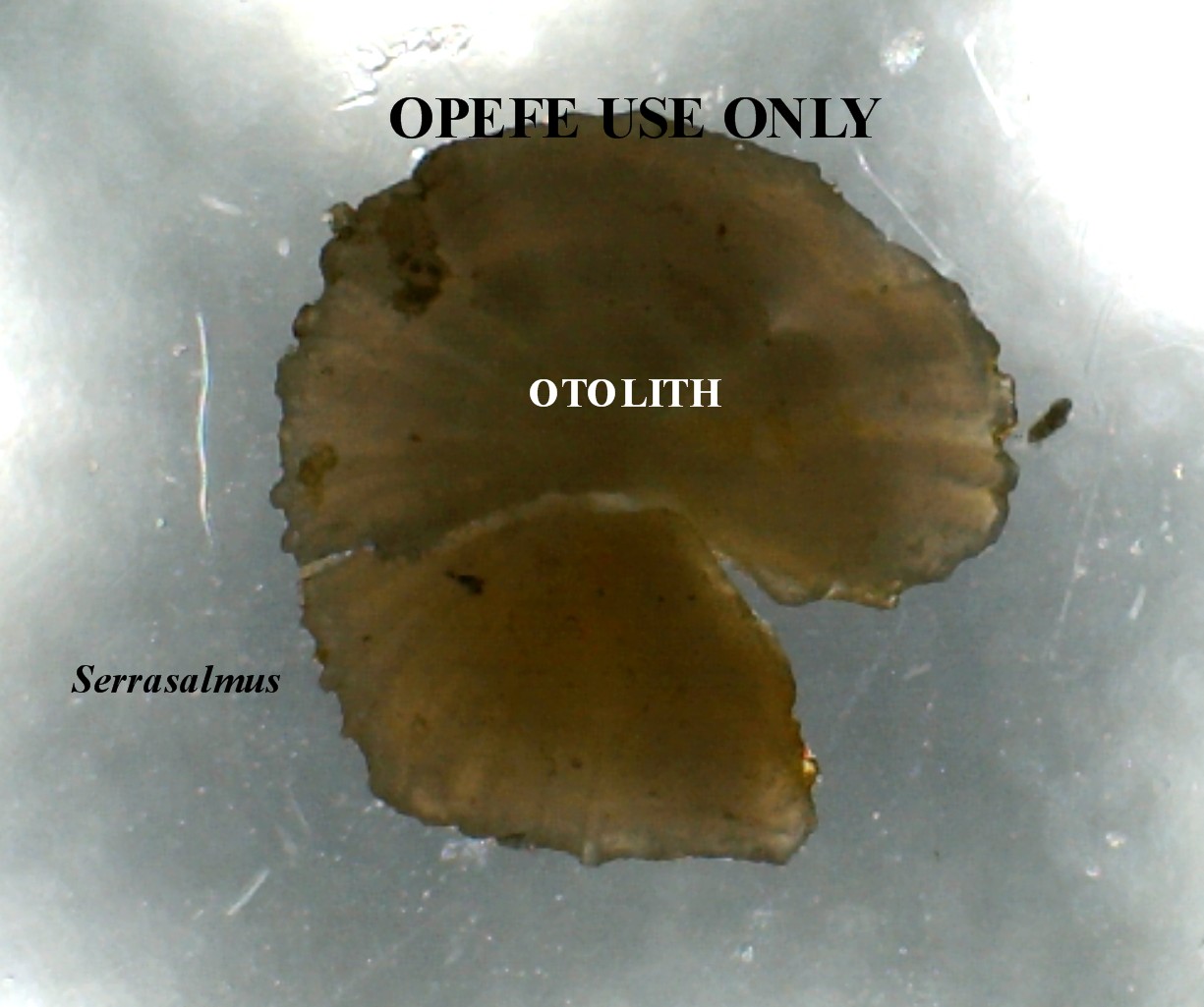
Determining a piranhas age (Otolith)
Use of scales was once the preferred method to determine fish age. The scale would be put under a microscope and the annuli (circle rings like a trees) would be counted. However, this method is now regarded as unreliable. Instead when the fish die, the bones in their heads called otoliths (oto meaning ear and lith meaning stone) are removed. These bones help the fish to keeping its balance in the water. When an otolith is removed from a fish, sectioned into thin slices and viewed through a microscope, it reveals a pattern of light and dark concentric rings. The only other way you can determine a piranhas age is to hatch it from an egg and record its growth.
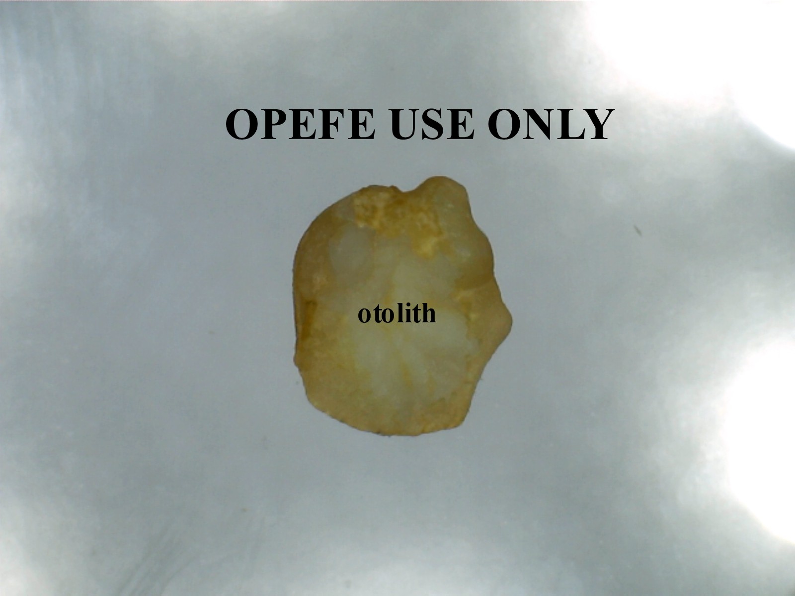
Unsliced otolith belonging to Pygocentrus nattereri |
Unsliced otolith belonging to Serrasalmus species. |
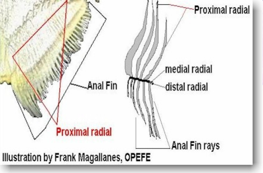
The fins
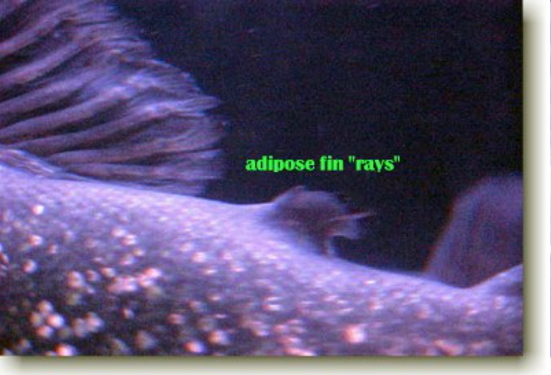 All piranhas possess fins which are used to
control their movements with the air bladder. The fins are paired and unpaired
and are named for the areas where they are inserted on the body. The only
exception being the adipose fin which is largely fatty tissue and this membrane
can possess rays or spines. All piranhas possess a predorsal spine (see photo
below) and combining all these features marks them all as members of the
Characidae family. P. piraya are unique in having rays in their
adipose fin. However, a few rare occasional examples of P nattereri
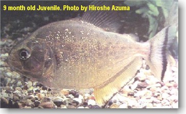
(southern portion - see photo top, left) have been known to have this feature.
Normally, however they are not rayed. The swimming motion is serpentine-like and
is done by the lateral body muscles. This motion is done in unison with the
caudal fin. Some authorities suggest the other fins are not of any real
importance in locomotion. The reason for this idea is because during fast
swimming, the fins are often folded close to the body. Their use largely is for
steering and act as a stabilizer. Often hobbyists will see the fish flick the
fins, this action is more or less a nervous action and can also act as a warning
to predators.
All piranhas possess fins which are used to
control their movements with the air bladder. The fins are paired and unpaired
and are named for the areas where they are inserted on the body. The only
exception being the adipose fin which is largely fatty tissue and this membrane
can possess rays or spines. All piranhas possess a predorsal spine (see photo
below) and combining all these features marks them all as members of the
Characidae family. P. piraya are unique in having rays in their
adipose fin. However, a few rare occasional examples of P nattereri
(southern portion - see photo top, left) have been known to have this feature.
Normally, however they are not rayed. The swimming motion is serpentine-like and
is done by the lateral body muscles. This motion is done in unison with the
caudal fin. Some authorities suggest the other fins are not of any real
importance in locomotion. The reason for this idea is because during fast
swimming, the fins are often folded close to the body. Their use largely is for
steering and act as a stabilizer. Often hobbyists will see the fish flick the
fins, this action is more or less a nervous action and can also act as a warning
to predators.
Predorsal spine
 The predorsal spine sits in front of the first spine
(ray) of the dorsal fin. This pre-dorsal spine is characteristic of all the
genera in Pygocentrus, Serrasalmus, Pristobrycon, and Pygopristis.
The predorsal spine sits in front of the first spine
(ray) of the dorsal fin. This pre-dorsal spine is characteristic of all the
genera in Pygocentrus, Serrasalmus, Pristobrycon, and Pygopristis.
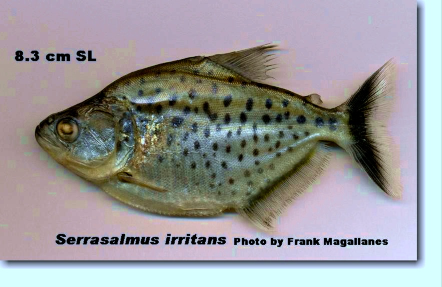
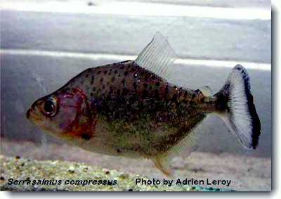
Tail markings
The caudal fin can take on different characteristics. They can all be very deeply prominent in having a black band or a band that is faded. It largely depends on the type of species, what type of damages the fin has suffered over the course of the fishes life and last, stress factors. Many of the Serrasalmus and Pristobrycon species have unique tail markings as shown below:
|
Serrasalmus irritans showing the dark "V" of the tail fin. |
Serrasalmus compressus showing the the black terminal band of the tail. Very common with majority of piranha and pirambeba species. |
Serrasalmus maculatus showing the subterminal band (or midway) on the tail fin. |
The hearing
Fish have ears, they lie entirely within the skull. They are paired, and consist
of membranous sac or sacculus. In piranhas, they have 3 utricles. These canals
lie in all three dimensions of space, two vertically, and one horizontally. In
bony fishes, the utricles is connected by means of the endoymphatic duct (a
small tube which is closed in bony fishes) which ends just below the skin in a
bulb, the endoymphatic sinus. The sacculus contains the ear stone, or otolith.
This bony concretion is secreted by delicate glandular tissue which is deposited
in proportion to the rate of growth for the fish. This makes it possible to
distinguish the age of fishes living in temperate and artic regions. The
internal part of the ear is filled with a liquid, the endolymph, whereas the
bony capsule surrounding the auditory organs contains perilymph.
The eyes
Vertebrates like snakes do not blink, but have a protective spectacle or brills. This is a transparent membrane in the skin which protects the cornea. Fish have a protective spectacle. Eyes of some teleosts (bony fishes) are protected by vertical folds of adipose tissue (fatty tissue) that may leave an aperture. One of those for example is the Hydrocynus sp. (African Tiger Fishes). When you see eye movement, like blinking motion it is probably the fish using a natural movement or exercise for whatever reason. Being in water, they don't need tear ducts like humans. The eyes are monocular which allows them to judge distances well. However fish are near-sighted. This may not be a handicap because even in clearest of waters it is difficult to see objects 100 feet away. The eyes of most fishes are located on the sides of the head. The eye function is like a photographic camera. This allows light to penetrate at different intensities which is transmitted to the optic lobes of the brain. The space between the the lens and retina is filled with the transparent liquid substance known as vitreous humor. The iris of the eye, capable of dilation and contraction, controls the amount of light passing through the pupil.
Sound
Helga Eichelberg
Zoological Institute, University of Bonn, Germany
Zoologisches Institut der Universität Bonn, Poppelsdorfer Schloß, D-5300 Bonn, Germany
21 September 1977
Summary The anterior and the posterior drum muscles of the piranha resemble each other in all essential fine structural aspects: myofibrils are slender; sarcomeres are short compared with those of other drum muscles; mitochondria, located in the periphery of the fibers, are numerous and show an irregular internal structure; and the sarcoplasmic reticulum is abundant. Triads appear at the level of the Z lines. The drum muscles have many motor endplates, which, however, lack the characteristic junctional-fold apparatus. No lipid substances could be demonstrated in these muscles. In the posterior drum muscle the fibers depart from their orderly longitudinal arrangement at irregular intervals.
In some species that have a hyaline humeral blotch, it is known that this region which harbors the air bladder effects the tympanum (produce sound) in some species. This sound resembles croaking or a buzzing sound depending on the species. When they are feeding it can be heard like a form of communication. Out of the water, large species can be a bit noisy and to the uninitiated, it can be frightening as well.
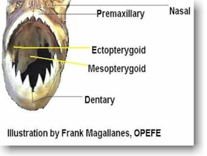
The teeth
|
|
|
|
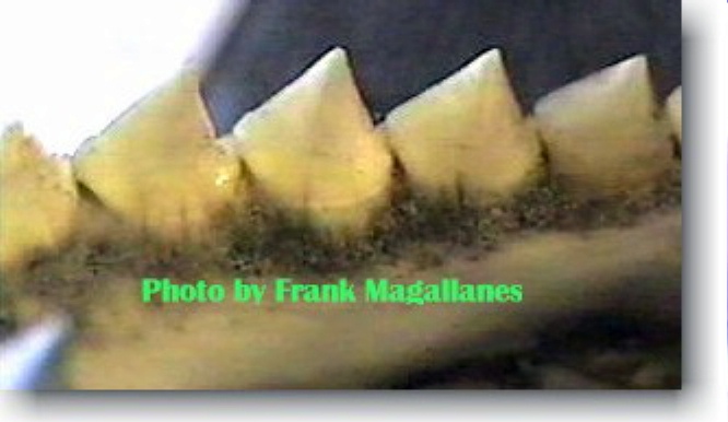
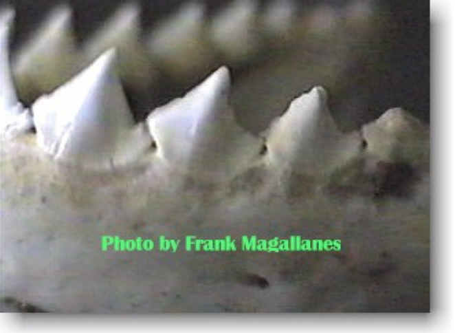
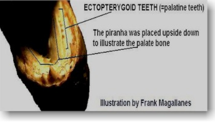
The images above show the formation of the teeth. The first image is the inside of the mouth and the second image is outside the mouth. Note the proximity of each tooth and how well formed they are. The lower jaw is massive and packs some rather large teeth as seen by the photo above. This projecting jaw gives the look of a bulldog in older specimens. The teeth are normally hidden from view by the thick lips. The tips of the teeth can be seen protruding on some species, while others they extend past the lips, giving the fish a large toothy appearance. The length of these teeth average 4 mm TL. The upper jaw, the teeth interlock with the lower and the palate region may carry some tiny bones or teeth-like structure called palatine or ectopterygoid teeth. These small teeth are not present in genus Pygocentrus, in Serrasalmus, these teeth may be worn down. Which is why historical authors placed these fish in genus Pygocentrus. The genus Pristobrycon these small teeth may or may not be present depending on the species.
The teeth of most piranha species are pointed and razor-sharp and tricuspid. Only the species P. denticulata is known to have teeth different from all other species of piranhas. The teeth of denticulata are crenulated (5 = penticuspid). Each piranha species have specialized teeth and the serration on the teeth vary.
Observations here at OPEFE dramatically show the P. denticulata species using them to shear off the husk of seeds.
For additional examples of piranha teeth click HERE.
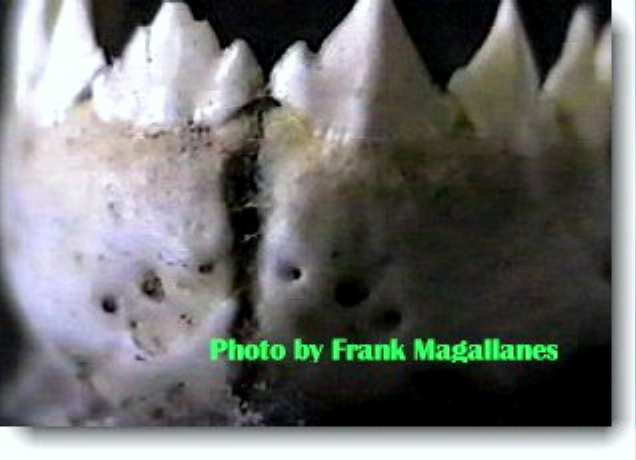
|
|
|
|

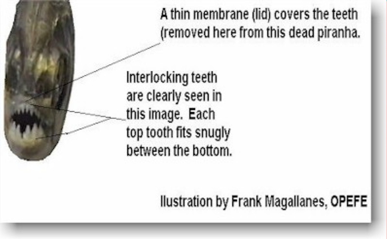
Each upper and lower tooth interlocks with each other (like a continuous saw blade). The teeth are not made to lacerate, crush, tear, or even used to hold on to prey fish. They are meant to clip off small pieces of flesh or fins (MYERS, 1949).
The lower jaw is part of a system of bones that form a structural arch called suspensorium, which is formed by the dentary, angular, articular, quadrate, sympletic,interhyal, meso and metapterygoid, hyomandibula and the opercular series. The jaw itself is formed by the dentary, angular and articular, but only the dentary carries teeth.
-
Neither the suborbital series nor cleithrum are part of the jaw system. The cleithrum is part of the scapular girdle, formed by the scapula, coracoid and cleithrum.
-
Linked to the jaw system are two other systems, the Hyoid complex and the branchial arch complex.
-
Teeth counts are hemi mandibules, 8+8 given that there is bilateral symmetry. (Pacus have symphysial teeth).
-
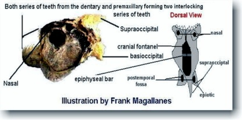
Both series of teeth from the dentary and premaxillar form two interlocking series.
|
This preserved specimen shows the teeth on the left of screen being replaced as a set. |
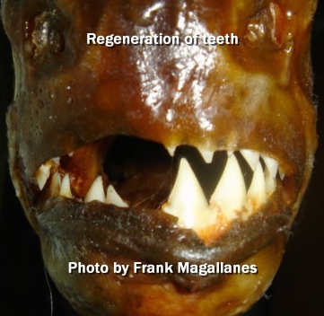
When a piranha loses a single tooth, it is replaced quickly as a whole set. This piranha is ready to lose its set of teeth. The new teeth are already formed in the gum area and do not drop down until the replacement is needed. Microscopic slides made at OPEFE do show the "ready teeth" on the standby position. Some of the individual species teeth are as variable as the species itself. Some are very broad while other are very narrow, and most are small for a species considered deadly. The largest tooth I have examined and measured based on a 23.5 cm SL specimen is only 8 mm (a bit larger than 1/4 inch long). The teeth are serrated which allows nice, clean slices. The piranha also have additional smaller teeth-like structures called "ectopterygoid teeth" (approximately 2-3 mm long). The ectopterygoid itself is a bone (palatine) is like an elongated plate that forms on the roof of the mouth with these small teeth.
These ectopterygoid teeth while useful for identification of species, can be worn with age and sometimes non-existent in older specimens like S. rhombeus. The lower jaw is actually two pieces held together at the center by tissue which acts like a glue. Only the genera Pygocentrus and Pygopristis lack these ectopterygoid teeth at all ages. The characins are the most heterodont fish group, that is, a group with very different teeth from one taxon to the other. The teeth are normally hidden behind the thick upper and lower lips. In few older examples of piranhas, particular when the set is about to be replaced the the tips of the teeth are exposed just above the lip line.
Exposed teeth
Piranhas teeth are normally hidden behind the thick lips only the points are partially exposed. At times, the teeth are completely exposed when a tooth is lost and the entire set is going to be replaced. Sometimes the lip is bit off during a feeding frenzy or when the fishes are in breeding condition. This gives the piranha a sinister appearance with teeth fully exposed. In South America, dead piranha lips are cut away to expose the teeth. These fish are then dried and mounted then sold as souvenirs to tourists. UNDER NO CIRCUMSTANCES CUT THE LIPS OFF A LIVE PIRANHA. Besides being unnatural, this is cruel and inhumane also this practice could cause the fish to become infected with a fungal disease. If you see a hobbyist do this to their fish, scold him. If you find a pet store doing this deliberately to their piranhas in order to enhance sales of their fishes, report them to the local Humane Society or local Aquarium Society. There are other organizations that also take complaints of animal cruelty seriously. While OPEFE is not associated with or affiliated with any particular animal rights group, sometimes getting their support, particularly when an animal is being harmed becomes a personal ethical choice.
The bite power
Dr. G. S. Myers, in The Piranha Book (TFH, M-539) gave the following Carl H. Eigenmann description:
"The best mechanical imitation of piranha teeth and jaws, and a very good imitation indeed, is a bear-trap, but one with teeth so sharpened on the edges, and the spring so strong, that they would clip off the bear's foot instead of merely holding it." No measurement of the biting power of a piranha has ever been made. It is my opinion the bite power is probably in the approximate range of 120-300 lbs sq. inch depending on size of fish. These estimates should not be considered as fact. Myers (1949) reported that "the power of the jaw muscles is such that there is scarcely living substance save the hardest ironwood that will not be clipped off." In 2011, a new study was done in the field using Serrasalmus rhombeus as a study specimen. The bite force was measured and it measured 70+ lbs. This is far greater bite than that of a great white shark of comparable size. (Steve Huskey, PhD). More to follow when the research is published pending 2012. READ THE NEWEST INFORMATION OF BITE POWER.
The intestinal tract
Piranhas are equipped by a spacious pharynx. The esophagous runs directly into the stomach without any indication between the two. A small constriction of the stomach marks where the small intestine begins which are looped or coiled like a hose and extends almost to the anus. It is this junction where the gonad tube meets into a thin tube that extends out to the anal opening. The rectum is barely discernable from this junction and intestine tube. Waste products are excreted from here. The liver is large and is composed of a high fat content. The fish does not possess a gallbladder.
The gills
The gills are used for respiration and the inside opening have gill openings and arches. These organs take up dissolved oxygen from the water. The gills are composed of branches which are reddish and very thin. They are highly veined and protected from the outside by the gill plate or operculum.
The lateral line
Running along the flank just behind the gill cover upper mid-body is the lateral line. This runs all the way to the distal end of the caudal peduncle. This lateral line is composed of a grooved, canal system that divides into three branches as it reaches the head. The lateral line is covered by scales and basically is a receptor of vibrations that enter the water. Hobbyists will sometimes notice their fish jump when a door is slammed or if foot steps are loud as they approach the aquarium. The fish senses these vibrations as they pass through the water to its lateral line. In all practical terms, it is a motion detector.
CONTRIBUTORS/ADVISERS:
-
Dr. Paulo Petry
-
Dr. Michel Jégu
REFERENCES
-
Myers, G. S., The Piranha Book, M-539, TFH, 1972
-
Miquelarena, Amalia Maria., Estudio Comparado del Esqueleto caudal en peces Characoideos de la Republica Argentina, (1981).
-
Weitzman, Stanley H., Osteology of Brycon Meeki (1962).
-
Fink, W. L. Revision of genus Pygocentrus 1993
-
Allometry and Developmental Integration of Body Form in a Piranha, Pygocentrus nattereri (Teleostei: Ostariophysi). Journal of Morphology 223:341-355 (1995)
USE YOUR BACKSPACE TO RETURN OR CLICK HERE TO RETURN RESEARCH PAGE
TO RETURN HOME CLICK HERE.
The OPEFE web site and its contents; is disclaimed for purposes of Zoological Nomenclature in accordance with the International Code of Zoological Nomenclature, Fourth Edition, Article 8.3 and 8.4. No new names or nomenclature changes are available from statements at this web site.
Copyright© 1994-2012 Oregon Piranha Exotic Fish Exhibit (The OPEFE fish exhibit is permanently CLOSED as of 2000) Sutherlin, Oregon. Information posted on this web site is archival data on fish scientific classifications and other information. DISCLAIMER: The copyrighted material may not be used for any purpose other than private study, scholarship or research. Cited information requires credit and this link www.opefe.com. All rights reserved. All images shown (unless otherwise noted) is property of OPEFE.